
Masseter
Masseter-Properties (revision)
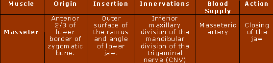
 The masseter is a quadrilateral muscle, there are two parts of this muscle the superficial and deep part. The superficial part is the largest part of the masseter muscle. This arises from thick tendinous aponeurosis from the zygomatic process of the maxilla and from the anterior 2/3 of the lower border of the zygomatic arch. The fibres in the superficial part of the masseter run diagonally and backwards. This structure is related to its function. The superficial portion of the masseter inserts into the angle and lower half of the ramus of the mandible.
The deep portion of the masseter is smaller and muscular in texture. This part of the muscle arises from the posterior 1/3 of the lower border, and the whole of the medial surface of the zygomatic arch. Insertion to the upper half of the ramus and lateral surface of coronoid process of the mandible.
Parotideomasseteric fascia (masseteric fascia) covers the masseter, which is firmly connected with it, this is a strong layer of fascia derived from the deep cervical fascia.
Origin/Insertion
The masseter is a powerful elevator of the mandible, which may be easily palpable when teeth are clenched. The broad origin of the masseter is related to its function, the most efficient function of this muscle includes unilateral chewing. Masseters are the strongest muscles of mastication in herbivores. In humans the masseter is the second most efficient muscle in mastication. The reason being the high ratio of neurons to muscle fibres is 1:600.
Image (top) courtesy of https://commons.wikimedia.org/wiki/Image:Muscle_masseter.png under the terms of the GNU free Documentation license.
Image (bottom) courtesy of https://www.flickr.com/photos/kchocho-travel-addict/851263630/ under the creative commons license.

The action of the masseter:
- Elevation of the mandible
- Lateral movements of mandible
- Unilateral chewing
- Retraction of mandible
The sole function of the masseter is to elevate the mandible (close the jaw) and crush objects between the molars, or in carnivores, to clench into other animals using the canine (fang) teeth.
Innervation
The masseter is supplied by the masseteric nerve which is a branch of the mandibular division of the trigeminal nerve (CNV) which runs over the mandibular notch. The masseteric nerve curves laterally above the lateral pterygoid to enter the deep surface of masseter.
Damage to the trigeminal nerve causes the patient to experience symptoms, like headache and migraine.